Significance: Treatment of unstable pelvic fractures relies on open reduction followed by internal fixation using surgical plates and screws, a surgery only performed by highly experienced and well-trained surgeons due to the complexity of the pelvic anatomy. The key challenge is the mental mapping of plate positioning and screw trajectories to the fractured anatomy in 3D, a skill that surgeons in training cannot easily acquire from conventional 2D training material such as textbooks, websites, or illustrations from their instructor. This suggests that an inherently 3-dimensional learning environment based on Augmented Reality (AR) has great potential to benefit surgeon training. Innovation and
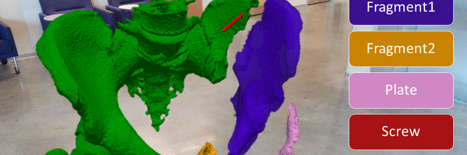
Approach: Our goal is to develop a collaborative and interactive AR learning platform targeted at improving the understanding of spatial relations of plates, screws, and anatomy during open reduction and internal fixation of pelvic fractures. Our system will be realized as an immersive 3D AR environment based on Microsoft’s HoloLens that visualizes virtual models of anatomy, plates, screws, and simulates the X-ray camera view to the student and instructor. It will allow for manipulation of every component to enable interactive investigation of optimal positioning of implants. This tool is designed to benefit anatomical education, as it will allow students to navigate around 3D structures promoting improved geometric understanding, increased knowledge retention, and a higher appreciation of function and structure.